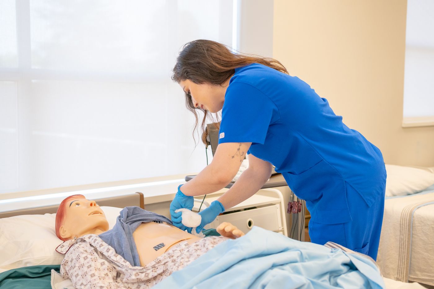
The tube passes without a struggle. The patient is drowsy and settles quickly. Nobody coughs. A syringe draws back a few millilitres of cloudy fluid that looks about right. A colleague pushes a little air down the tube and hears that familiar gurgle over the stomach. The tube is secured, the feed is hung, the pump is started, and the nurse moves on to the next room feeling that the job went well.
Every single one of those observations is also compatible with a tube sitting in the right main bronchus.
That is the whole argument of this article, and it is worth sitting with before reading further. The dangerous moment in nasogastric tube care is not the insertion. A tube entering the airway during insertion is a recognised, recoverable event; the tube comes out and the attempt is reconsidered. What is not recoverable is the decision made in the minute afterwards, when someone concludes the tube is in the stomach on the basis of evidence that never proved anything, and then feeds, flushes or medicates through it. Liquid delivered into a lung is the harm. The verification step is the only thing standing between the two.
This piece is about that step alone. It describes what verification is, how it is taught and how it is assessed, not how to perform an insertion. Its narrower job is to remove five specific habits from a reader's mental toolkit, because each of them has been mistaken for confirmation for decades and none of them ever was.
Most curricula introduce the nose to earlobe to xiphoid measurement, usually shortened to NEX. The tube is laid along a path from the tip of the nose, to the earlobe, and then down to the xiphoid process, and the resulting length is noted as an estimate of how much tube is likely to be needed to reach the stomach.
That estimate is genuinely useful for what it is. It gives a defensible starting figure instead of a guess, and it makes the difference between an adult and a smaller adult visible before anything is touched. It has also been criticised in the nutrition literature for a specific reason: the surface path it traces tends to fall short of the true internal distance, so NEX has a known tendency to underestimate the length required to place the tip well within the stomach rather than at or just past the gastro-oesophageal junction. Some programmes therefore teach a corrected version of the measurement.
None of that repairs the fundamental limitation. NEX produces a number, and a number is a scalar. It describes distance along a path that has not yet been chosen. The oesophagus and the trachea begin from the same pharynx and will both accept a tube of the same length. A perfectly measured, perfectly corrected NEX is exactly as consistent with a tube in the bronchial tree as with a tube in the stomach. The measurement is silent on the only question that matters, which is not how far but where.
The distinction that carries everything. Measurement answers "how much tube". Verification answers "which tube did it go down". They are different questions, and no amount of precision on the first one produces an answer to the second.
The habits below are still in circulation, still demonstrated informally on wards, and still offered by candidates in skills validation sessions as their evidence of correct placement. Each fails for a reason worth understanding, because the reason is what stops the habit returning under pressure.
Treating NEX as confirmation is the error described above, applied in reverse. The tube reached the expected marking, therefore it is in the expected place. The logic only holds if the tube can be in exactly one place at that depth, and it cannot. Length agreement is a check that the tube has not been left far too short or pushed far too deep. It is not a location.
Air is pushed down the tube while a stethoscope is held over the epigastrium, and a gurgle or whoosh is taken as proof the tube is in the stomach. The mechanism of failure is acoustic. The abdomen is not an isolated sound chamber, and a stethoscope reports only that a sound reached the diaphragm, not where in the chest or abdomen that sound was generated. Air delivered into a tube whose tip lies in the oesophagus, or in the bronchial tree, transmits perfectly well through the intervening tissue and is heard in the same place, with much the same character. The test produces a positive result in every scenario it was supposed to tell apart.
The same problem in its general form. The American Association of Critical-Care Nurses, in its practice alert on initial and ongoing verification of feeding tube placement in adults, states plainly that auscultation is not useful for distinguishing respiratory from gastrointestinal placement. That is the sentence to carry: not that auscultation is imperfect, or that it should be combined with something else, but that it does not discriminate between the two situations a verification method exists to separate. A method that returns the same signal for both answers has no diagnostic content, however experienced the ear.
Coughing during passage is often described as the body's own alarm for airway entry, and when it happens it is informative. Its absence is not. A cough is a reflex with prerequisites: intact sensory pathways in the larynx and trachea, a level of consciousness sufficient to mount the response, and the muscular capacity to generate the effort. Sedation blunts it. Obtunded or reduced consciousness blunts it. Neurological injury, bulbar involvement and existing artificial airways can abolish it altogether. Precisely the patients most likely to need enteral access are the patients least able to produce the warning. Silence is a statement about the reflex, not about the tube.
Fluid returns up the tube, it is cloudy or off-white or faintly green, and it is accepted as gastric. Respiratory secretions, however, occupy the same visual range. They can be clear, cloudy, whitish, tinged or thickened, and pulmonary fluid drawn back through a narrow tube does not announce its origin. Colour and consistency overlap between the two compartments, which means visual inspection can raise suspicion but can never exclude airway placement. The eye is a screening impression, not a test.
What all five share. Each one gives the same result whether the tube is in the stomach or in the airway. A test that cannot come out differently in the two situations it is meant to distinguish is not a test. It is a ritual that produces confidence without producing information, and confidence is the specific thing that gets a feed started.
It is easy to read the list above and conclude that the people who taught these methods were careless. They were not. The nasogastric tube is an old device, and for most of its history there was no bedside chemical test within reach and no imaging available on a general ward in the small hours. Clinicians had a stethoscope, a syringe and a patient who needed feeding. The air bolus was a reasonable improvisation for its era, and it was transmitted the way most bedside practice has always been transmitted, by demonstration from one shift to the next rather than by evidence.
Watch on YouTube: The Wild History of the Nasogastric Tube!, from our educator's own channel, Spice to health$Nursevibes.
That history explains the persistence. A practice that begins as the best available option acquires the authority of habit, and habit does not automatically retire when a better option arrives. The whoosh test kept its place because it is fast, free, needs no order and no waiting, and delivers an immediate, memorable, sensory result. It feels like evidence. Everything about the experience of performing it reinforces the belief that something has been checked.
Which is exactly why it is dangerous rather than merely useless. A method that produced no feeling of certainty would have been abandoned quietly. This one produces certainty reliably and accuracy not at all, and the certainty is the ingredient that gets a pump switched on.
Against that background, the position of the professional bodies is unusually consistent. The American Association of Critical-Care Nurses, in the practice alert already cited, directs that a radiograph visualising the entire course of the tube, interpreted by a clinician competent to read it, is obtained before a newly placed tube is used for the first time. The phrase "entire course" is doing deliberate work: a film showing only the tip in an expected region does not exclude a tube that has taken an unexpected route to get there.
Testing the pH of aspirate is the other method taught almost universally as a bedside check, and it works on a real physiological difference rather than a sensory impression, which is why it survives where auscultation does not. But there is a point here that must not be glossed over, and it is the most important sentence in this article: the pH value that counts as acceptable is set by the reader's own facility protocol, and it is not the same everywhere. Guidance in the United States and guidance in the United Kingdom do not use an identical figure, individual employers set their own threshold within their policy, and a number printed on a public webpage would therefore be the wrong number for a large share of the people reading it. The threshold that applies to any given nurse is the one written in the policy of the place where that nurse practises, and nowhere else.
The American Society for Parenteral and Enteral Nutrition addresses the same territory in its safe practice recommendations for enteral nutrition therapy, and it has gone further than restating the rule. Its NOVEL project exists specifically to examine new methods of verifying enteral tube location, which is a candid acknowledgement from within the profession that the current bedside repertoire is imperfect and that better tools are actively being sought. That is worth knowing, because it frames the present guidance as the best available answer rather than a settled one.
In the United Kingdom the framing is blunter still. NHS England, continuing work begun by the former National Patient Safety Agency, treats a misplaced nasogastric tube used for feeding as a never event. The British Association for Parenteral and Enteral Nutrition works in the same space. The never event category is not a synonym for "rare"; it is a statement that the occurrence is considered wholly preventable when existing safeguards are applied. The classification is an argument about the verification step, not about bad luck.
There is a second, quieter failure mode, and it catches experienced nurses more often than new ones. A tube that was confirmed this morning is treated as a confirmed tube for the rest of its life. It is not. Confirmation describes where the tube was at the moment it was checked, and tubes move.
The list of things that move them is mundane, which is the problem. Vomiting and retching can bring a tube back up into the oesophagus or further. Vigorous coughing shifts it. Suctioning, repositioning in bed, sitting a patient forward, transfers to and from a trolley, restlessness and a confused patient's own hand all act on a tube that is held in place by a piece of tape. The AACN practice alert covers ongoing verification for exactly this reason, and it treats it as a continuing obligation rather than a single event at insertion.
This is why the documented external length matters so much and why it is more than paperwork. A recorded number at the nose is the only cheap way a change in position becomes visible to the next person, on the next shift, who did not see the tube go in. Without it, migration is invisible until something goes wrong. This is the same discipline that underpins any serial observation, and it is worth reading alongside our guide to the head-to-toe assessment, where the value of a documented baseline is the identical argument in a different setting.
Everything above collapses into one operating rule that is short enough to survive a busy shift: nothing is fed, medicated, flushed or connected to suction through a nasogastric tube until its placement has been confirmed to the standard the facility sets.
The flush deserves particular attention, because it is where the rule is most often quietly broken. A flush feels like a preliminary rather than an administration, a small volume of water that hardly counts. It counts. Water delivered into a lung is water in a lung regardless of the intention behind it, and in practice the flush is very often the first thing that goes down a newly placed tube. Treating it as exempt removes the protection at precisely the moment the protection is needed.
The one-line version. An unconfirmed tube is not a tube with a small doubt attached. It is a tube whose destination is unknown, and an unknown destination is treated the same way as a known airway until evidence says otherwise.
The corollary is documentation and handover. A tube whose position has not been confirmed is recorded as unconfirmed and handed over as unconfirmed, so that the status travels with the patient rather than dissolving at shift change. Most of the reported harm in this area involves someone acting on an assumption inherited from a colleague, which is a communication failure wearing the costume of a clinical one.
In a supervised skills session, the insertion itself is the part learners worry about and the verification block is the part that decides the outcome. Candidates who handle the tube confidently frequently stumble at the moment they are asked what evidence they have that the tube is where they believe it is, because the honest answer often turns out to be one of the five habits above, absorbed on a ward and never examined.
That is a fixable gap, and it is fixed by rehearsal rather than by reading. Our four-hour assessed NG tube course is built around exactly this asymmetry: it is capped at six learners so every person is observed individually, it costs $149, and the validation checklist is published in full on the course page so nobody arrives guessing what they will be assessed against. Most people who do not pass it do not fail at insertion. They fail after the tube is already in.
For readers preparing for licensure rather than for a skills validation, the same content appears in examinable form, and the question writers tend to probe the same weak point by offering an attractive distractor built from an old habit. Our free set of NCLEX-style IV and NG practice questions with rationales is a useful way to find out whether the distinction between measuring and verifying is genuinely secure or merely familiar.
Educational use. This article is learning material for nurses and nursing students. It is not clinical advice, and it does not replace your employer's policies, your facility's protocols, or the judgement of a licensed clinician. Always follow the standards and procedures in force where you practise.
No, and describing it as a backup is what keeps it alive. A supplementary test is only worth adding if it can change the conclusion, and the air bolus cannot: it produces an audible result whether the tip is in the stomach, the oesophagus or the bronchial tree. Adding it to a proper verification adds nothing but time, and using it when a proper verification has not been completed adds false confidence, which is worse than no check at all.
Deliberately not stated here. The acceptable threshold is a bedside parameter set by each facility's own protocol, and guidance in the United States and the United Kingdom does not use an identical figure. A number published on a webpage would be wrong for a large proportion of readers, and being confidently wrong about this particular number is exactly the failure the article is about. The applicable value is the one written in the policy where a nurse practises.
It cannot. Respiratory secretions and gastric fluid overlap substantially in colour and consistency, so an aspirate that looks gastric is consistent with a correctly placed tube and also consistent with a tube in the airway. Visual inspection can raise suspicion and prompt further checking, but it cannot exclude the outcome that matters.
Ongoing verification is part of the standard, and the American Association of Critical-Care Nurses practice alert addresses it explicitly alongside initial verification. The reason is mechanical rather than theoretical: tubes migrate after vomiting, retching, coughing, suctioning and repositioning. The method used and the intervals required are set by the local protocol, which is where those specifics belong.
The clinical principle is shared, but the regulatory packaging is not. NHS England, building on work from the former National Patient Safety Agency, classifies a misplaced nasogastric tube used for feeding as a never event, a category that carries specific reporting and investigation consequences. The American Association of Critical-Care Nurses and the American Society for Parenteral and Enteral Nutrition express the same expectation through practice alerts and safe practice recommendations instead. Different instruments, the same underlying rule.
Reading about verification changes what a nurse believes. Being observed doing it, and being asked out loud what evidence supports the conclusion, is what changes what a nurse does at two in the morning with a feed waiting and a bay full of patients. That is the gap our NG tube session is designed to close, with six learners, a published checklist and an assessor watching the verification block rather than the insertion.