Photo: Carly Dernetz via Pexels
Every unit has the patient nobody wants to start a line on. Years of chemotherapy, dialysis, dehydration or simply their anatomy mean nothing is visible and nothing is palpable, and the day's plan stalls while three people try, apologize, and try again. The patient knows the script better than the staff do.
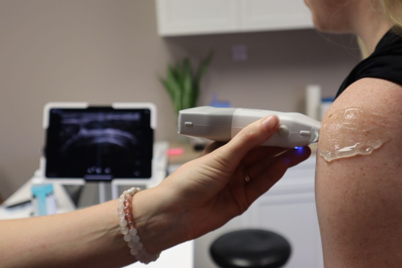
Ultrasound guidance changed that. A probe on the arm shows veins that cannot be seen or felt, and a clinician trained to use it reaches them on the first attempt far more often than one working by touch. The evidence is consistent enough that the standards governing infusion practice treat visualization technology as part of the plan for difficult access rather than a specialty extra.
This article covers the skill as a program teaches and assesses it: why difficult access exists, what the standards say, what the trials found, what a training program contains, who may perform it, and what equipment familiarization means. It does not teach the technique, which is learned on a phantom and then on patients with an instructor beside you.
Peripheral IV insertion is the most frequently performed invasive procedure in healthcare, and it fails more than most people realize. Helm and colleagues (Journal of Infusion Nursing, 2015) put peripheral catheter failure at 35 to 50 percent and titled the paper "Accepted but Unacceptable." The 2024 consensus from the Association for Vascular Access (AVA), written with the Infusion Nurses Society, the American Association of Critical-Care Nurses and ECRI, repeats those figures and defines difficult intravenous access, or DIVA, as a patient who has experienced multiple unsuccessful insertion attempts, listing extremes of age, female sex and veins with limited visibility or palpability among its correlates.
The cost has been measured. Witting (Journal of Emergency Medicine, 2012) followed adults needing an IV in an urban emergency department and graded difficulty by what it took to succeed. Sixty-one percent were cannulated with one puncture. Eleven percent needed more than one puncture. Twenty-three percent needed a second non-physician clinician, with a median delay of 15 minutes. Five percent needed a physician, with a median delay of two hours. A history of difficult access made the two harder groups 2.5 times as likely.
The Infusion Nurses Society's Infusion Therapy Standards of Practice, ninth edition (Journal of Infusion Nursing, 2024), is the document most US infusion policies are written against. It carries a standard on vascular visualization, and the position across recent editions has been consistent: technology such as ultrasound is used to increase insertion success in patients with difficult venous access, and clinicians who use it are educated in it and demonstrate competency first. The exact wording belongs to the Standards, which your education department should hold.
The AVA consensus of 2024, endorsed by the Emergency Nurses Association, APIC and a dozen other bodies, turns that into organizational duties. Organizations develop policies for ultrasound-guided peripheral insertion, including aseptic non touch technique and disinfection of the equipment after use. Every inserter is trained in DIVA assessment and in when to escalate. Clinicians are trained and demonstrate competency in the facility-approved visualization technology. And organizations establish a clear escalation pathway for failed attempts or when no vein is visible or palpable.
For nurses specifically, the Emergency Nurses Association's General Assembly resolved in 2011 to support ultrasound-guided peripheral venous access within the registered nurse scope of practice, on the basis that emergency nurses can perform it safely after appropriate education and training, and the ENA now publishes a clinical practice guideline on difficult intravenous access.
Printable study guide
Get the printable IV Insertion Training reference
IV Insertion Training from Wahero Health Institute is a printable reference on the standard color-coding system for identifying cannula size at a glance, a quick companion for the didactic session of any vascular access program.
Pay by Cash App, PayPal or Zelle at checkout; the download link is emailed once the payment is confirmed, usually the same day. Study material only: it does not replace the Board's requirements, your employer's policies or the judgment of a licensed clinician.
The outcome that matters most to the patient is whether the first attempt works, and here the literature is clearest. Egan and colleagues (Emergency Medicine Journal, 2013) pooled seven randomized trials in difficult access and found a pooled odds ratio of 2.42 for successful cannulation with ultrasound; Stolz and colleagues (Journal of Vascular Access, 2015) pooled seven studies and reported 3.96. Van Loon and colleagues (British Journal of Anaesthesia, 2018) analyzed eight studies and 1,660 patients across all levels of difficulty: success was 81 percent with ultrasound against 70 percent without, an odds ratio of 2.49, with fewer punctures, less time, higher satisfaction and no reduction in complications. Tran and colleagues (Journal of Emergency Nursing, 2022) pooled seven randomized studies of nurse-performed cannulation, 527 patients, and found ultrasound roughly doubled the odds of first-attempt success (2.08) while attempts, time and satisfaction were similar.
One result cuts the other way. The Cochrane review by Tada and colleagues (2022), covering 16 trials and 2,267 participants, found ultrasound probably increased first-pass success in difficult access (risk ratio 1.50, low-certainty evidence) but decreased it, and probably increased pain, in patients whose veins were easy. Ultrasound is a tool for the identified difficult patient, not a default, and assessors expect candidates to say so.
The evidence in one sentence. In difficult access, ultrasound roughly doubles the odds that the first attempt works; in easy access it does not help and may hurt. The competency includes knowing which patient is in front of you.
Published nurse programs share a four-part shape: didactics, phantom practice, supervised insertions, and sign-off with ongoing review. Feinsmith and colleagues (Journal of Emergency Nursing, 2018) ran a four-hour course for emergency nurses with competency defined as ten successful supervised insertions; of 34 enrolled, 12 reached it within nine months, their success rose from 81 percent in their first ten attempts to 96 percent in attempts 21 to 30, and difficult-access attempts fell 7 percent. McKinley and colleagues (Cureus, 2024) trained 16 emergency nurses with one hour of didactics, one hour on a live model and a homemade simulator, then supervised shifts until each reached ten successes: 200 attempts, 80 percent success, 14 of 16 credentialed, and a median of 11 attempts to reach ten.
Three details matter. The phantom stage builds hand-eye coordination cheaply: McKinley's group used a homemade simulator, which fits our argument about what simulation fidelity actually buys. The supervised stage is where attrition happens, because it needs a competent instructor with protected time; Feinsmith's 35 percent completion was a staffing result, not a learning one. And the commonest reason an attempt failed in the 2024 study was losing track of the needle on the screen, which tells a program where its phantom hours should go.
No national certificate makes a nurse eligible for ultrasound-guided insertion. Eligibility comes from the state's scope-of-practice framework and the employer's policy; the AVA consensus lists nurses, respiratory therapists, paramedics and medical providers among those who insert peripheral catheters, consistent with state law.
In Texas, the Board of Nursing's Scope of Practice Decision-Making Model, which replaced the older six-step model, asks eight questions in order: whether the activity is prohibited by the Nursing Practice Act or Board rules, whether a valid order or protocol authorizes it, whether it fits facility policy, whether it aligns with evidence and national nursing organization positions, whether the nurse has documented competence, whether a reasonable and prudent nurse would do it, whether the nurse can accept accountability, and, for LVNs, whether supervision is available. A stop at any step puts the activity outside scope; documented competence is the question this article is about.
Facility policy adds the operational layer: which units, which patients, how competency is recorded and reassessed, and how the catheter is documented and cared for, which is why infiltration and extravasation assessment sits in the same policy.
Familiarization is a knowledge domain of its own, usually assessed in the didactic session. Candidates are expected to identify the high-frequency linear transducer used for superficial vessels and say why, to know what depth and gain do to the image and where the vascular preset lives, and to state what the facility requires for probe covers, gel and cleaning. Catheter selection for the vein found is assessed alongside it, and our printable IV Insertion Training reference covers the cannula color-coding at a glance. None of this is the psychomotor skill; it is the vocabulary that lets an instructor coach you.
A sign-off is an observed insertion against a checklist, not a count on its own. Assessors typically score patient identification and consent, DIVA assessment and the decision to use ultrasound, machine setup and probe preparation, aseptic technique, the insertion, confirmation on screen that the catheter is in the vein, securement and dressing, documentation that records the device, the site and the use of ultrasound, and the handling of a failed attempt, including when to stop and whom to call. Knowing when to stop is a standard, not a preference, as our article on what IV insertion competency really takes argues, and the competency includes knowing your facility's escalation pathway.
What a sign-off is not. Competency decays without use, and the published programs all built in review. Ask what your facility's reassessment interval is before you ask for the probe.
Educational use. This article is learning material for nurses and nursing students. It is not clinical advice, and it does not replace your employer's policies, your facility's protocols, or the judgement of a licensed clinician. Always follow the standards and procedures in force where you practise.
Can a registered nurse place ultrasound-guided peripheral IVs? In many US facilities, yes, after a training program and documented competency, under a policy the organization has written. The Emergency Nurses Association's 2011 resolution supported it within the registered nurse scope of practice after appropriate education and training, and the 2024 AVA consensus expects trained, competency-assessed clinicians. Whether it applies to you depends on your state board's position and your employer's policy; in Texas the Board of Nursing's Scope of Practice Decision-Making Model is the tool.
Does ultrasound guidance help every patient? The evidence says no. The 2022 Cochrane review by Tada and colleagues found ultrasound probably increased first-pass success in difficult access but decreased it, and probably increased pain, in patients with easy veins. Standards therefore frame ultrasound as a tool for identified difficult access, not a default for everyone.
Does this article teach the insertion technique? No. Needle-to-probe technique is a supervised psychomotor skill learned on a phantom and then on patients with an instructor beside you, using your facility's equipment and policy. This article covers why the skill exists, what the evidence says, what a program contains and how competency is assessed, which is the part that transfers between settings.
A one-day class produces nurses who have held a probe. A program produces nurses who are signed off, documented, reassessed and backed by a policy. Wahero Health Institute delivers vascular access training on site in Texas, from didactics and phantom practice through supervised competency validation against your own policy.
Educational content only
This material is published by Wahero Health Institute for professional education and is not individual medical advice, a care protocol, or a substitute for clinical judgment. Always follow your facility's policies, your state's nurse practice act, and your own scope of practice, and confirm medication doses against a current authoritative reference before administration. See our Terms of Use.