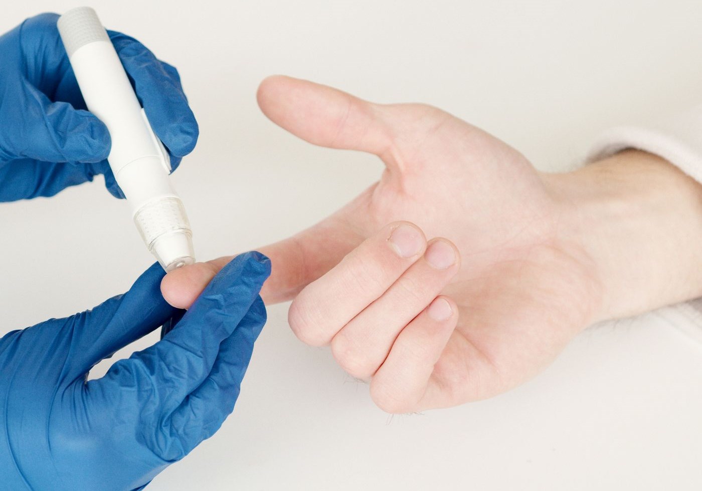
A 62-year-old man is two days out from a bowel resection. No history of diabetes, no steroids on his chart, no dextrose running, and a normal A1c from his pre-admission workup. His capillary glucose comes back at 214 mg/dL. Nothing on the medication record explains it, and the number is not falling.
What the chart cannot account for, physiology can. His adrenal glands are producing cortisol at several times their resting output, and cortisol's metabolic purpose is to put glucose into the blood and keep it there. This is not a laboratory error, and it is not the sudden onset of diabetes. It is the signature of an activated stress response doing exactly what it evolved to do.
Understanding that mechanism changes how you read a glucose result. This article covers the HPA axis and cortisol's daily rhythm, the four routes by which cortisol pushes glucose up, why acutely unwell patients run high without diabetes, the pattern steroid courses produce, what it all means for nurses who work nights, and how Cushing syndrome shows the same physiology at full volume.
Video: Cortisol and Diabetes, from our own educator's YouTube channel, Spice to health$Nursevibes.
Cortisol arrives at the end of a three-stage relay called the hypothalamic-pituitary-adrenal axis, or HPA axis, and knowing the stages tells you where things can go wrong.
A stressor, whether sepsis, surgery, trauma, hypoglycemia, pain or fear, is registered by the hypothalamus, which secretes corticotropin-releasing hormone (CRH). CRH travels a short portal circulation to the anterior pituitary, which releases adrenocorticotropic hormone (ACTH). ACTH reaches the adrenal cortex and drives the zona fasciculata to synthesize cortisol from cholesterol. The sequence takes minutes, not seconds, which is why cortisol is the slower, sustained arm of the stress response while adrenaline handles the first heartbeat of it.
The axis also switches itself off. Circulating cortisol feeds back negatively on the hypothalamus and pituitary, suppressing CRH and ACTH. That one loop explains a great deal of clinical medicine: it is why a patient on long-term exogenous steroids cannot mount a stress response of their own, and why abrupt discontinuation after prolonged therapy is treated so cautiously.
Cortisol is also one of the body's principal timekeeping signals, released in pulses that follow a daily pattern set by the suprachiasmatic nucleus of the hypothalamus. Levels reach their lowest point around the middle of the night, climb in the small hours, and peak shortly after waking, a surge known as the cortisol awakening response. From there they decline across the day to the overnight trough.
That rhythm has a metabolic job: the morning peak mobilises fuel for the transition from sleeping to moving, which is one reason fasting glucose can sit slightly higher on waking than it did at two in the morning. Whenever a rhythm is that consistent, its absence becomes informative. Loss of the normal nocturnal trough is one feature endocrinologists look for when investigating cortisol excess, and late-night salivary cortisol testing exists precisely because a healthy axis should be quiet at that hour.
Cortisol is a glucocorticoid, and the name is a job description: a steroid that raises glucose. It does so through four complementary routes, and each one shows up somewhere on a patient's chart.
Hepatic gluconeogenesis. Cortisol upregulates the liver enzymes that build glucose from non-carbohydrate precursors, including phosphoenolpyruvate carboxykinase and glucose-6-phosphatase. The liver becomes a glucose factory running independently of what the patient has eaten, which is why a nil-by-mouth patient can still run high.
Substrate supply. A factory needs raw material. Cortisol promotes proteolysis in skeletal muscle, releasing amino acids, and lipolysis in adipose tissue, releasing glycerol and free fatty acids. Those substrates feed gluconeogenesis, and the same catabolic effect explains the muscle wasting of prolonged glucocorticoid excess.
Peripheral insulin resistance. Cortisol interferes with insulin signaling beyond the receptor in muscle and fat, reducing recruitment of GLUT4 transporters to the cell membrane, so less glucose leaves the blood for a given amount of insulin. Free fatty acids from lipolysis worsen the same resistance, and the two effects compound.
Permissive action on other hormones. Cortisol amplifies catecholamines and glucagon, the other counterregulatory hormones, while adrenaline blunts insulin release from the pancreatic beta cell. The acutely stressed patient therefore has more glucose entering the circulation, less leaving it, and a weakened insulin brake on both.
Put those mechanisms into an acutely unwell patient and stress hyperglycemia is the predictable result. Sepsis, myocardial infarction, major surgery, burns, trauma and severe pain all activate the HPA axis, and the elevated glucose that follows is a physiological response rather than a new disease.
The American Diabetes Association describes this in its Standards of Care as hyperglycemia occurring in hospitalized patients without a prior diagnosis of diabetes, using a glucose above 140 mg/dL as the threshold that prompts attention, and it recommends an A1c to distinguish previously undiagnosed diabetes from a purely stress-driven rise. That distinction matters after discharge: an A1c in the diabetic range says the glucose problem preceded the admission, while a normal A1c points to the stress response and to a patient who nonetheless deserves follow-up screening.
What nurses observe is a glucose that tracks the severity of the illness, rising as the patient deteriorates and settling as they recover. The same logic we apply to recognizing patient deterioration early applies here: a single glucose value is a snapshot, and the direction of travel across a shift carries more information than any one reading. Note the trend, note what else changed at the same time, and document both.
One caution belongs with this. Physiology is not permission to explain away a number. If the pattern does not fit the clinical picture, that is a reason to reassess and escalate, not to reason around it, and hospital glycemic management is governed by your facility's protocol rather than by a rule of thumb.
Everything above applies with more force when the glucocorticoid comes from a pharmacy rather than an adrenal gland. Therapeutic steroids are prescribed at doses that far exceed physiological cortisol output, and steroid-induced hyperglycemia is a well-recognized consequence in patients with and without pre-existing diabetes.
The pattern has a shape, and the shape follows the drug's duration of action. With an intermediate-acting glucocorticoid such as prednisone given as a single morning dose, the glycemic effect builds through the day: fasting morning glucose is often the least affected value, while readings climb through the afternoon and peak in the late afternoon or evening before easing overnight. Longer-acting agents such as dexamethasone spread the effect across the full 24 hours and blunt that pattern.
The teaching point for nurses is observation, not intervention. A patient monitored only at breakfast on a morning steroid dose can look deceptively well controlled while running high every afternoon, which is why monitoring schedules on steroid therapy are usually written to capture the later part of the day. Document the timing of each reading against the dosing time, and the record becomes interpretable to the next clinician instead of ambiguous. Expect the pattern to move when the therapy moves: dose changes, tapers and switches between agents all shift the curve. Our educator's video above walks through the same cortisol and glucose relationship if you prefer to hear it explained once.
This physiology is not confined to your patients. Cortisol's rhythm is anchored to the light-dark cycle, and night shifts ask the body to be alert when the axis is programmed to be quiet, then to sleep when it is programmed to peak. Circadian misalignment of this kind is associated with a flattened, phase-shifted cortisol profile rather than the crisp curve in the figure above.
Sleep restriction pushes in the same direction. Controlled laboratory studies of shortened sleep have repeatedly reported reduced insulin sensitivity in healthy adults, alongside changes in appetite-regulating hormones. Combine that with meals at 03:00, limited access to real food and caffeine to stay sharp, and the metabolic risk profile of shift-working nurses becomes an occupational health question rather than a lifestyle lecture.
Serious bodies treat it that way. The International Agency for Research on Cancer classifies night shift work involving circadian disruption as probably carcinogenic to humans. The American Nurses Association has published a position statement on addressing nurse fatigue, framing it as a shared responsibility of employers and nurses rather than individual willpower. NIOSH offers free training for nurses on shift work and long work hours, a better starting point than most generic sleep advice.
None of that makes night work avoidable. It does mean that anchoring sleep timing, protecting a real sleep block after nights, controlling light exposure and eating on a schedule are physiological interventions rather than wellness slogans, which is why they come up in our live training sessions with new graduate cohorts.
If stress hyperglycemia is cortisol excess for days, Cushing syndrome is cortisol excess for months to years, and every feature of it maps back to the four mechanisms above.
Sustained gluconeogenesis and insulin resistance produce glucose intolerance and, in many patients, frank diabetes. Sustained proteolysis produces proximal muscle weakness, thin skin and poor wound healing. Fat redistribution produces the characteristic central obesity with relatively thin limbs. Add hypertension and the loss of the normal diurnal rhythm, and the clinical picture is the figure earlier in this article held down for a year.
Two points are worth carrying away. First, the most common cause of Cushing syndrome overall is not a tumour but prescribed glucocorticoid therapy, which is why a careful medication history matters. Second, the Endocrine Society's clinical practice guideline on diagnosing Cushing's syndrome describes tests, including late-night salivary cortisol and dexamethasone suppression testing, that are direct applications of the rhythm and the feedback loop above. Diagnosis sits with the medical team; the nurse contributes accurate observation and an understanding of why those tests are ordered.
Can stress alone cause a high blood glucose in someone without diabetes? Yes. Sepsis, surgery, trauma or myocardial infarction activates the HPA axis, and the resulting cortisol and catecholamine surge raises glucose by increasing hepatic production and reducing peripheral uptake. The ADA calls this stress hyperglycemia and recommends an A1c to determine whether diabetes was already present.
Why is my patient's glucose high when they have not eaten anything? Because cortisol makes glucose rather than absorbing it. Hepatic gluconeogenesis runs on amino acids and glycerol released from muscle and fat, so the supply is internal. Nil-by-mouth status does not switch off the stress response.
What glucose pattern should I expect on a morning dose of prednisone? Typically the fasting value is least affected, with glucose climbing through the afternoon and peaking in the late afternoon or evening. Longer-acting agents such as dexamethasone spread the effect across the whole day. Your facility's protocol sets the monitoring schedule.
Does working nights really affect my own blood sugar? Circadian misalignment flattens and shifts the cortisol rhythm, and controlled sleep restriction studies have repeatedly reported reduced insulin sensitivity in healthy adults. IARC classifies night shift work involving circadian disruption as probably carcinogenic, and the ANA treats nurse fatigue as a shared employer and nurse responsibility.
How does Cushing syndrome relate to everyday steroid therapy? Same physiology, different dose and duration. Prescribed glucocorticoids are its most common cause overall, which is why medication history matters and why prolonged therapy is not stopped abruptly.
Endocrine physiology stops being memorisation the moment the mechanism clicks, and everything from steroid side effects to NCLEX endocrine questions gets easier afterwards. Wahero Health Institute teaches this material in live online sessions with time for questions, and enrolled learners revisit the recordings, notes and practice questions through the student portal. For the bedside skills that go with the theory, our simulation lab puts them in your hands.
Educational content only
This material is published by Wahero Health Institute for professional education and is not individual medical advice, a care protocol, or a substitute for clinical judgment. Always follow your facility's policies, your state's nurse practice act, and your own scope of practice, and confirm medication doses against a current authoritative reference before administration. See our Terms of Use.